UWL anatomy lab uses human cadavers to aid in student learning
March 4, 2020
According to Thomas Greiner, Associate Professor of anatomy in the health professions Department at the University of Wisconsin-La Crosse, UWL has 18 cadavers on campus: 14 in the Health Science Center and four in Prairie Springs Science Center.
“All the University of Wisconsin systems that make use of donated bodies, obtain them through the donation program that is run at the medical school in Madison,” said Greiner. “We are the second-largest anatomy program in the state that Madison supplies.”
Many different departments make use of the donated cadavers, studying the body from head-to-toe.
“The anatomy laboratory is used primarily by students in physical therapy, occupational therapy [OT], physician assistant, and the CRNA Programs but also is used by exercise sports science, and biology as well,” said Greiner.
According to Greiner, UWL keeps the cadavers for approximately six months before sending them back to Madison.
“We get the cadavers at the start of the summer program and then exchange some of the cadavers at the end of the summer with new ones,” said Greiner, “So when the OT anatomy programs take classes in the Fall, they dissect the new cadavers, as well as observe the previously dissected ones.”
After UWL returns the cadavers to Madison, they are cremated and returned to families, if desired.
“The people that donate their bodies can indicate whether they want their remains returned to their families for burial or if they’ve got no desire to have things returned, I sometimes take specimens off them to keep and show students,” said Greiner. “When the bodies get returned, they are all cremated in Madison and the ashes are returned to the family if desired, if not they are buried in a secret place.”
The cadavers are honored in different ways while on campus in efforts to respect those who chose to donate their bodies and aid UWL students’ education.
“I tell my students since they are all going to be clinicians, ‘Treat them like they are your first patient.’ So we don’t do anything that is outside the realm of our learning goals and one of the things that I constantly remind my students is we have to protect the modesty of our patients to respect them,” said Greiner. “We honor the gift by doing a full and complete dissection and learning as much as we can, making sure none of their gift is wasted.”
“We also have a memorial service in the Spring honoring the donors and their families,” said Greiner.
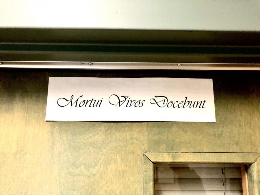
The sign above the door to the anatomy lab reads, “Mortui vivos docebunt, which means, “The Dead Teach the Living,” which Greiner said is to be a valuable part of students’ educations.
“What students really get out of the process is the understanding of no two people look alike,” said Greiner. “We look around us and we intuitively know that but yet because we have an Anatomy atlas that has only one set of pictures and we expect everyone to look like that on the inside, but they don’t and are different.”
“So I can either tell students that, or I can make them experience that,” said Greiner.
“It [anatomy lab] has given me a lot of hands-on experience to really see what the different sections of the body actually look like,” said Emily Garfoot, a sophomore biomedical science major who uses an anatomy lab weekly.
Greiner finds the use of cadavers to be an experience that students cannot achieve by using textbooks or computer programs.
“A clinician could learn all the ways of treating and examining a patient through book learning and computer models, while never having touched a single patient until they are actually out there in the clinic. Would you want a clinician like that?” said Greiner. “They could probably do that, take a written test and do just as well, but that doesn’t mean they learned how to deal with a human patient at a really intimate level, and that cannot be replaced.”
“I honestly do not know how I could have as much knowledge about the human body as I do if I did not have access to cadavers,” said Amelia Thompson, a sophomore biology major with a pre-med track at UWL. “Lab manuals are very helpful and do have images of cadavers but having the hands-on experience of being able to trace the arterial and venous system or feel the difference between the semimembranosus and the semitendinosus helps make learning easier.”
“I think it is important to have the cadavers because it gives students the ability to not only look at models and photos of anatomical structures, but it also gives them the experience to interact with an actual cadaver which is something not a lot of people have done in their lives,” said Garfoot. “I feel like it helps our anatomy classes really excel at teaching the students.”
“If you talk to any clinician out there, they will have stories about their first anatomy lab and the will remember back on what the cadavers taught them,” said Greiner.
“I believe that having cadavers available for study is incredibly beneficial to students. By having the ability to familiarize myself with the human body, I have found that my knowledge of the human anatomy has stuck in my brain long after I learned it,” said Thompson. “We are lucky to have access to cadavers and I have great respect for those who donated their bodies so students like me can receive a better, more in-depth education.”
To learn more about anatomy labs at UWL visit, https://www.uwlax.edu/grad/physical-therapy/gross-anatomy-laboratory/.


